46 Wimpole Street, London
020 7490 7222 Book an appointment
020 7490 7222 Book an appointment
Ophthalmic surgeons over the last century or more have come up with all sorts of interesting and novel ways of improving vision for patients. This article discusses just some of these techniques, including the excimer laser that we use to correct vision today. Most of these techniques have fallen by the wayside, including one of the first most common methods known as radial keratotomy.
The First Step
Radial keratotomy was first put forward in the early 1900’s by an Ophthalmologist called Lans, and then interest was rekindled in the 1930’s by Professor Sato in Japan where he was using this technique to treat a corneal condition called keratoconus. The whole concept was to flatten the central cornea. To do this he would make small incisions in the cornea from the endothelial side (the back of the cornea). Radial keratotomy was then popularised far more in the 1970’s and 1980’s by Professor Fiodorov in Moscow. 10 years later it was taken up by the United States.
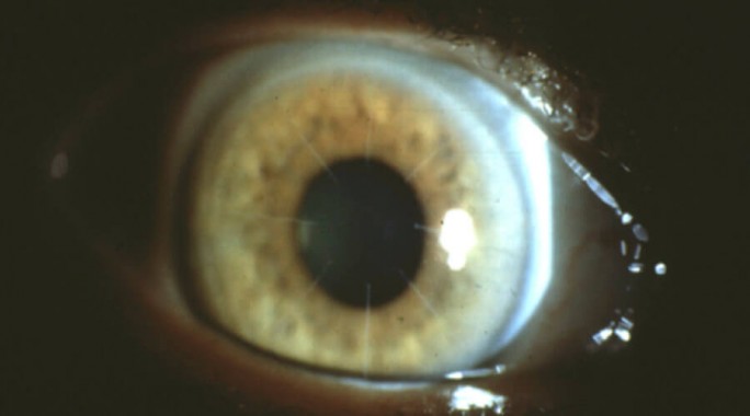
Radial keratotomy showing the characteristic incisions on the cornea.
The main problem with radial keratotomy was that when incisions or cuts were placed into the cornea they produced fine scars. However carefully created the scars are, they can cause some corneal damage which then caused glare and scattering of light especially at night when the pupil gets a bit larger. The other problem is one of predictability, and the fact that a lot of patients treated would have their short sight corrected quite well but then find in the years to come they became more long-sighted, something termed a ‘hyperopic drift’.
Introduction of Lasers
Lasers were first invented in the early 1960’s based upon the work of Albert Einstein from the 1920’s. The 50th anniversary of laser was just recently celebrated in May 2011. There had been numerous developments and applications of lasers during that period, but ophthalmology was the very first speciality in medicine to adopt them.
They were first used for treating diabetic retinopathy (a complication of diabetes, caused by high blood sugar levels damaging the back of the eye – the retina) with an Argon laser and there have since been numerous ophthalmic applications, the most exciting of which is the excimer laser used in laser eye surgery which was first put forward in the 1980’s.
The difference between using an excimer laser and radial keratotomy is that the entire cornea was being reshaped, rather than using individual cuts. It was really more of a surface treatment, so you weren’t doing something as invasive in the cornea and because it was under computer control, the altered prescription would be very precise. Our initial treatments involved treating damaged parts of the eye in order to remove corneal scarring or clouding, but then we moved on to treating short-sight by altering the shape of the cornea.
In order to correct short-sight with laser refractive surgery, we have to effectively flatten the cornea slightly. This first procedure was known as Photorefractive Keratectomy (PRK), and I carried out the very first PRK procedure in the UK in November 1989 at St Thomas’ Hospital, London.

Early scanning electron micrographs from David Gartry’s doctoral thesis of a PRK in a donor cornea. The amphitheater appearance is created by the laser operating through an expanding iris diaphragm aperture (just like in a camera) with more tissue removed centrally and progressively less as the edge of the donor cornea is approached. This is a basic myopic PRK therefore in which the corneal profile has been flattened – the cornea is less steep and the light rays reach the retina.
The initial problem with PRK in the early days was that the first prototype lasers had various technical constraints and restrictions, one of the main restrictions being that we had to use small diameter treatment zones which meant we couldn’t treat the whole of the pupil area. This resulted with patients that were overall very happy with their vision but unfortunately could have problems when their pupils became larger at night time. Working on the surface of the cornea with these very early prototype lasers meant that patients would often have very variable results.
Some patients would tend to generate haze in the cornea from PRK which could sometimes cause a reduction in the level of vision or contrast sensitivity. This corneal haze would tend to cause the patient to go back to being short-sighted, which we call ‘regression’. It would also tend to cause night vision problems.
In the early days we couldn’t treat astigmatism, we couldn’t treat hyperopia (long-sight) and we had no tracking which meant that we couldn’t be sure that we were treating exactly in the middle of the pupil, so if the patient moved during the treatment they could potentially end up with a suboptimal result. Thanks to new tracking technology, this is no longer an issue.
The LASIK Revolution
In order to try and get around the problem of having corneal haze, Professor Palikaris from Crete suggested that we do the laser surgery inside the cornea by carrying out a technique called LASIK (Laser Assisted in-situ keratomiluesis). To get inside the cornea we had to create a very thin layer, which was called the corneal flap. This flap allowed us to carry out the treatment within the cornea. The very fact of having a relatively smooth surface meant that we hardly ever saw corneal haze again. The results were more predictable, there was less regression and we could even treat hyperopia and higher prescriptions. LASIK is now the treatment of choice for most patients seeking laser eye surgery.
In the early 2000’s, the concept of wavefront technology was introduced. Newspapers started to advertise what they termed ‘Super Vision’ because in theory if you can carry out a wavefront guided treatment, then you can get rid of a patient’s short-sight, astigmatism and any other optical imperfections resulting in perfect vision. Wavefront treatments have become routine nowadays.
Bubble Technology
The next big step was to generate the corneal flaps using femtosecond lasers instead of using a mechanical bladed microkeratome. This was a major step forward because the flaps could now be created with tremendous precision. We could program the exact diameter and the exact depth of the flap in a very accurate way as compared with the microkeratome.
Femtosecond lasers are now also used in cataract surgery. The modern era of cataract surgery really began in the early 90’s, with phacoemulsification technology which meant that we could replace the human lens with very small incisions. The benefit of now having part of the surgery carried with a laser means it is very precise. When we perform lens replacement surgery, we now have the option of putting in premium lenses such as toric lenses to correct astigmatism and multifocal lenses to correct both near and far distances. This is a great treatment option for patients who either aren’t suitable for laser eye surgery or who need a more permanent solution to needing reading glasses.
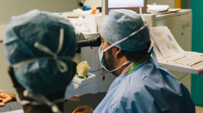
Modern laser eye surgery is now safer, faster and more accurate than ever. David Gartry is pictured here using the STAR S4 Excimer laser to correct a patient’s vision. The procedure is over in minutes.
Sophisticated Imaging
Other technological advances have come along in tandem with laser eye surgery, and we’re much better now at analysing the cornea than ever before. Technology such as the Pentacam Scheimpflug system provides detailed measurements of corneal thickness and 3 dimensional scanning of the front part of the eye, which means that we can eliminate patients from surgery who are unfortunately not suitable due to their cornea being too thin or mis-shapen. Another development in corneal imaging is Optical Coherence Tomography (OCT). It provides a cross-section of the cornea, enabling you to measure the thickness of a corneal flap, even 20 years after surgery!
Laser eye surgery is now safer, faster and produces better results than ever before. Our patients often refer to it as life-changing and indeed the freedom from glasses or contact lenses can bring newfound experiences to many. If you would like a laser eye surgery consultation, please do get in touch with one of our friendly team. We look forward to seeing you.